Microplastic "A threat to human life"
Image
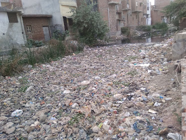
     Plastics: A life threatening problem Plastic is one of the life threatening problem of world. Now, the situation is out of the neck on garbage management issue.  Microplastics in drinking water :- Microplastics are every where in our environment it have been detected in marine water, wastewater, fresh water , food air and drinking water, both bottled and tap water. Microplastics enter freshwater environment in a number of ways :-  Image 1 primarily from surface run-off and wastewater effluent (both treated and untreated),  but also from combined sewer overflows,  industrial effluent degraded plastic waste and atmospheric deposition. The limited evidence show that some microplastics found in drinking water may come from treatment and distribution system for tap water and bottling or bottled water.

Lung Disease Detection by Sound Analysis

   Detection of lung Disease by lung sound analysis                                                    technique

There are some steps by which can detect the disease which can arise in lung by only using the sound produced from lung. But there are some major problem arise during the processing of these sound which are as:-
  1. separation of Noise from the sound sample.
  2. separation of heart sound from the lung sound.
  3. To get the lung sound database. 
Now there are the required steps through which we can easily detect the lung disease by the analysis of sound 

To understand the lung sound analysis there are two required slides which can enhance the knowledge about the sound produced by lung.


Slide No.1 :- Lung sound characteristics


Slide No.2:- Waveforms of Sound which show the disease indications.


Now we can start how can we detect the lung disease by analyzing the sound of lung

Slide No.3:- Location of the microphone on the posterior lung. 


Slide No. 4:- Receiving of sound from different sources.


Slide No.5:- Extraction of Noise from the sound sample.


Slide No.6:- Processing of  Noise free sound in computer in line.

Slide No.7:- Separation of lung sound and heart sound from the sample.

Slide No.8:- Demonstration of sample after separation of sound we make the wave-graph of only lung sound.


 Know after making these wave graph these wave graph divided into 20 fragments and after fragmentation these sound sample we are shown into time expanded image and then these graph are compared with the Slide no.2.


Comments

Related blogs

How to lose Fat and obesity??

Application of NCBI?

Microplastic "A threat to human life"